Media
HKU’s finding on avian influenza A (H7N9) virus well adapted to infect human respiratory tract reveals why numerous human infected cases in a short period of time
25 Jul 2013
HKU research team reveals why the avian influenza (H7N9) virus led to over 130 human cases in a relatively short period of time. By using human respiratory tract tissues maintained in culture, the researchers discover that the avian influenza A (H7N9) virus is as efficient as the 2009 pandemic H1N1 virus, commonly known as swine influenza, in infecting the human respiratory tract. Compared to the avian influenza A (H5N1) virus, H7N9 virus might pose an even more important pandemic threat to human. The research has just been published online today in the international scientific journal, The Lancet Respiratory Medicine.
This research was led by Professor Malik Peiris, Tam Wah-Ching Professor in Medical Science and Chair Professor of Virology, School of Public Health and Professor John Nicholls, Clinical Professor of The Department of Pathology, The University of Hong Kong Li Ka Shing Faculty of Medicine. Professor Malik Peiris gives remarks, “The research team has made use of a human respiratory tract ex vivo explant culture system to show that the H7N9 viruses are better adapted to infect and replicate in the human respiratory tract than other avian influenza viruses. The ability of avian influenza viruses to infect the human airways (trachea, bronchus) determines how readily these viruses can jump from birds to humans, to assess the adaptability of the virus in human and to potentially adapt to human-to-human transmission. Hence the findings are crucial for the future research.”
Research findings
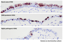
The researchers used human respiratory tract tissues (bronchus and lung) maintained in culture to compare infection with the avian influenza A (H7N9) virus, the 2009 pandemic influenza A (H1N1) virus (commonly known as swine influenza) and highly pathogenic avian influenza A (H5N1) virus. They find that the H7N9 virus infects and replicates in both human bronchus as well as the H1N1 virus, and far more efficiently than the H5N1 virus.
These studies also identify the type II alveolar epithelial cells within the lung as a key target for H7N9 virus replication. This is the key cell type supporting regeneration and repair of damaged lung tissues. Thus infection and damage to these cells will prevent the repair processes that allow the lung to recover from injury or infection.
Taken together, this study demonstrates that the H7N9 viruses are better adapted to infect and replicate in the human airways and pose an important pandemic threat, perhaps even more so than H5N1 virus.
Research background
Since February 2013, there have been 134 laboratory-confirmed human influenza A (H7N9) virus infections reported. There have been 43 deaths so far. The source of human infection appears to be poultry. There is so far no evidence of sustainable human-to-human transmission within the community.
About the research team
Researchers at the School of Public Health, the Centre of Influenza Research, the Department of Pathology and the State Key Laboratory of Emerging Infectious Diseases of The University of Hong Kong Li Ka Shing Faculty of Medicine, led the research. Other research team members included Dr Michael Chan Chi-wai, Assistant Professor, Dr Renee Chan Wan-yi, Research Assistant Professor, Ms Chan Lok-yung, MPhil student, Dr Chris Mok Ka-pun, post-doctoral fellow, Dr Leo Poon Lit-man, Associate Professor and Professor Yi Guan, Daniel CK Yu Professor in Virology, from the Centre of Influenza Research and the School of Public Health, HKU Li Ka Shing Faculty of Medicine.
Please visit the website at http://www.med.hku.hk/v1/news-and-events/press-releases/ for press photos.
The image shows human ex vivo bronchus culture, research team compared the extent of virus infection with the avian influenza A (H7N9) virus, the 2009 pandemic influenza A (H1N1) virus (commonly known as swine influenza) and highly pathogenic avian influenza A (H5N1) virus in the human bronchus tissues cultured ex vivo.
The part with brown color shows virus infected cells and the blue indicates non infected cells.
H7N9 virus leads to extensive infection of the surface cells (epithelium) of the bronchus, comparable to the 2009 pandemic virus. In contrast, the H5N1 virus only infects occasional bronchial epithelial (surface) cells.

